Dr Graham Treece, the newly appointed Evelyn Trust Lecturer in Engineering for Clinical Practice, is organising a Joint Workshop on Image Processing in Medicine to strengthen links with those involved in this field. The workshop is aiming to bring together anyone in Cambridge with an interest in image processing as applied to medicine.
The workshop will be split into four sessions, covering major themes in image processing as it applies to medicine across a range of image acquisition techniques and physical scales from molecules through cells, tissues and organs:
- Date: Monday 17 November 2008
- Time: 09:15 - 17:35
- Venue: The Cancer Research Institute on the Addenbrooke’s site.
The workshop has already attracted 30 speakers from 18 university departments, with over 100 people currently registered. But there are a few more spaces for anyone interested in this field from the Engineering Department. Registration instructions can be found at http://talks.cam.ac.uk/talk/index/13407
- Morphology and Heterogeneity will cover the measurement and analysis of shape and composition.
- Imaging Mechanical Properties will address topics such as tissue stiffness and compliance.
- Analysing Motion / Change will cover the measurement of flow and techniques for motion tracking.
- Novel / Enhanced imaging will cover techniques for increasing spatial and functional dimensionality.
Each session will consist of longer talks providing an overview to introduce the relevance of the session in Medical applications, and very short talks from members of the local research community introducing some of their own work relating to each theme.
All talks will be at a fairly high level, suitable for a broad audience, concentrating on what has been achieved, what still needs to be achieved, and why, rather than going into technical details. Each session will be closed with a discussion, aiming to pull together common research strands across discipline boundaries and potential future research directions.
This workshop is being jointly organised by:
Engineering for Clinical Practice
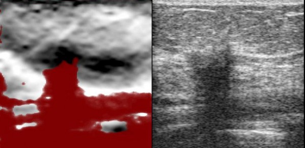
Dr Graham Treece's own research covers novel uses of ultrasound in medicine. The figure above shows both a conventional ultrasound image (on the right) and an ultrasound strain image or 'elastogram' (on the left). The strain image reveals stiff (dark) and soft (bright) regions, as well as indicating areas of poor data quality (red). These images both come from the same raw data - but the strain image reveals information which is impossible to see in the normal ultrasound image.
Tumours are often stiffer than surrounding tissue and these can show up very well using strain imaging. In the example, the patient has two stiff (black) regions - an invasive carcinoma of the breast (centre), but also a ductal carcinoma in situ (right), which is only visible on the strain image.
Graham has been involved in engineering / clinical collaborations for ten years, and has commercialisation experience through licensing of software and patenting of novel algorithms.
