A new CT (Computer Tomography) machine has been installed in the Materials Lab, which is already finding multiple uses ranging from examining the effectiveness of needle-less syringes to the fracture behaviour of Kevlar re-inforced concrete.
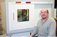
A technique more commonly used for medical applications, computer tomography is used to produce three dimensional images from multiple X-ray readings.
Alan Heaver explains:
"This equipment was built to our specification by X-Tek, initially so that we could examine the internal failure mechanisms of metallic foams. The X-rays reveal components having different densities. We have a large specimen chamber and we are developing techniques for in-situ dynamic testing. These tests can be observed in real time using the X-Ray source, and we can build up a complete three dimensional model of a specimen using a series of images. When the model is complete, it is possible to filter the information and look at components of particular density. This is a very powerful technique, which could also be used for imaging circuitry inside an electronic package for instance.
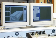
The example shown above (left) shows a concrete sample with a Kevlar fibre running through it. The CT image next to it (centre) has been processed so that details of the fibre running through the sample, the voids in the concrete and the plastic casing are all that can be seen. The right hand image is of an electronic component. The X-ray image reveals details of the circuitry within the plastic casing.
