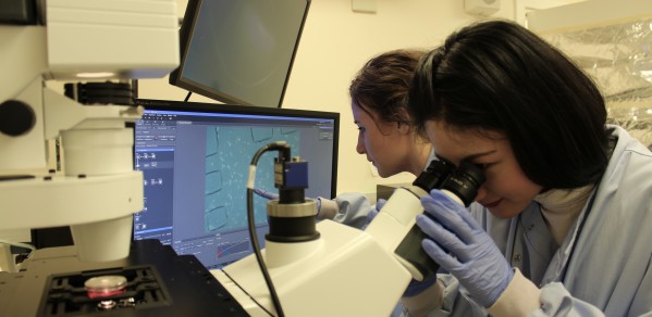
Bioscientist attitudes towards the future of microfluidics-based 3D culture technology and the search for their ‘ideal’ model have been revealed in a new research survey conducted by the Department of Engineering.
We hope that the results of our survey can be used as a type of ‘quality check’ with regards to the functionality of 3D culture options and as a means of encouraging standardisation to be a pathway for regulatory change.
Dr Huang
Organ-on-a-chip and vasculature-on-a-chip are examples of microfluidics-based 3D cell/tissue culture models which are created with microchip manufacturing methods that arrange living cells to simulate tissues and organs.
But a new research survey from the point of view of the end users – the biomedical community (including industrial scientists)1 – has shown that a ‘significant gap’ exists between the desired systems and the existing 3D culture options.
The research, led by Dr Yan Yan Shery Huang, Lecturer in Bioengineering, revealed that those surveyed (42) had expressed concern that the reliability, functionality and validation of engineered tissues may prevent the uptake of new cell culture technologies, but that these obstacles should be overcome by future 3D models.
Microfluidics often require specific training, ‘in-house recipes’ and can involve complicated external setups such as syringe pumps. The challenge, therefore, lies in simplifying user operation, while ensuring that future microfluidics continue to create possibilities for tissue engineering and next generation drug testing.
When questioned about their usage of 3D culture systems, such as microfluidic chips, the majority of the researchers surveyed did not have previous experience (only 7.1% were familiar with this technique). However, 85.7% of the researchers expressed a clear interest in adopting microfluidic culture, either performing it by themselves (50%) or with collaborators (35.7%).
But the more complex the system, the greater the time needed for system installation and associated training needs. Of the researchers surveyed, 61.9% indicated they would like to see the completion of the preliminary training and setting up of a 3D culture platform within four weeks.
The survey showed that advanced-career researchers (18 out of 42), with more than seven years of experience in their fields, have higher expectations for the complexity, physiological relevance of a 3D culture system, and expressed interest in customisable products.
Dr Huang and her co-authors Elisabeth Gill and Ye Liu believe this stark contrast with the preferences of early-career researchers, points to the need for greater consideration in the design and marketing of 3D culture systems.
“The growth of microfluidics-based 3D culture technology has rapidly emerged over the past two decades, with the ultimate aim to boost the development of fundamental bioscience and pharmaceutics,” said Dr Huang.
“Interesting microfluidic models have been engineered, such as organ- and vasculature-on-a-chip, but microfluidics are not without their weaknesses. Our survey is a first example to take a look at the potential use of microfluidic cell culture technology among biomedical researchers, at a time when there is a clear drive towards more predictive models based on human derived cells rather than animal tests. This is because the latter are not suited as a reference model for validation. Although the number of researchers surveyed was limited, this still provides a glimpse into the expectations of the biological community.
“We hope that the results of our survey can be used as a type of ‘quality check’ with regards to the functionality of 3D culture options and as a means of encouraging standardisation to be a pathway for regulatory change.
"In order to achieve the ideal 3D culture model and broader uptake, we believe a more ‘killer application’ may need to be showcased; one which achieves the level of ‘baseline’ complexity that the biomedical researchers desire, as well as improved system reproducibility, functionality and user friendliness. Any hesitation to do so may be a hurdle for new culture systems to be accepted as valuable tools for fundamental biomedical research and drug screening in an industrial context.”
It is hoped the survey findings may provide academic insight for entrepreneurs interested in the commercialisation of these systems.
Microfluidic on-chip biomimicry for 3D cell culture: a fit-for-purpose investigation from the end user standpoint Future Science OA: DOI:10.4155/fsoa-2016-0084
1 A total of 42 researchers completed the questionnaire. They include 15 research group leaders, 24 lab-based researchers and three industrial scientists. Their research covers areas including cancer, neuroscience, stem cell, toxicology, endocrinology and ageing.

