From non-invasive glucose monitoring to rapid food and materials analysis, a new class of optical spectrometer could transform how and where high-precision measurements are made.
Traumatic Brain Injury needs to be urgently prioritised on the world health agenda, argue research groups from the Departments of Engineering and Clinical Neurosciences.
Dr Lengwe Sinkala (International Health Systems Group at Cambridge) will research how paediatric surgical services can be redesigned to better support neurodivergent children.
Uni spin-out, CamGraPhIC, receives EC greenlight for €211m funding (about £183m) from Italy to support the development of photonic optical transceivers based on graphene.
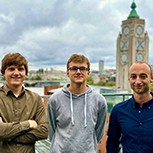
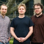
DropCode founded by three Cambridge alumni, is accelerating bioengineering with a droplet-microfluidics platform that uses DNA barcodes to run millions of experiments faster and cheaper.
Differences in cellular pathway activity flip the switch from nocturnality to diurnality and explain a major evolutionary change humans have undergone.