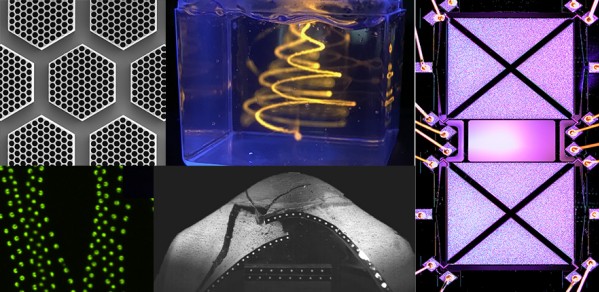
Striking imagery and video that encourages you to look at engineering through an artistic lens has won its creators prizes in the Department’s 2020-21 Photography Competition, sponsored by ZEISS (Scanning electron microscopy division).
The five prize-winning entries that caught the judges’ attention can be seen in the slideshow below. They include:
- A microscopic image of carbon nanotubes
- An image showing embedded 3D printing in action
- High-speed camera footage of the collapse of thin mortar scale-model vaults
- A microscopic image of droplets captured during the development of new methods of drug release
- A microscopic image of two MEMS (micro-electro-mechanical systems) resonators.

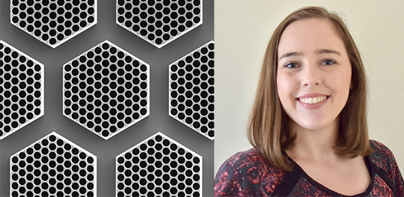
First Prize
Awarded to PhD student Aoife Gregg, who is part of the EPSRC in Nanoscience and Nanotechnology (NanoDTC) at the University of Cambridge, for her image titled Honeycombs of carbon nanotubes. Aoife is supervised by Professor Michaël De Volder.
“My image, taken using a scanning electron microscope, shows forests of vertically-aligned carbon nanotubes which have been grown into the shape of hexagonal pillars with inner pores in a honeycomb pattern,” she said. “Carbon nanotubes are already used commercially to add strength and conductivity to composite materials, and further applications range from sensing to microelectronics to battery electrodes. The carbon nanotubes shown here will be combined with a polymer to drive the honeycomb cells to reversibly close and open, depending on their environment. The hexagonal pillars in this image are 450 microns wide, but they can easily be scaled up or down depending on the desired application.”
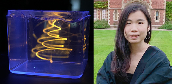
Second Prize
Awarded to Iek Man Lei for her image titled A suspended Christmas tree. At the time of entering the competition, Iek was a PhD student in the Biointerface Research Group at Cambridge, supervised by Dr Yan Yan Shery Huang.
Iek said: “My photo shows a suspended ‘Christmas tree’ printed in a support bath. The experiment was performed with a laboratory-built 3D robotic bioprinter that I developed during my PhD. The setup enables fabrication of soft materials, transforming them into 3D functional structures. In the experiment here, a pluronic ink (a synthetic polymer) was printed in a Carbopol support bath that exhibits a yield stress property. The pluronic ink was stained with rhodamine B, an orange-red fluorescent dye.
“Embedded 3D printing is an emerging additive manufacturing technology that enables creation of freeform and overhanging structures of soft materials, such as hydrogels and elastomers. During the embedded printing process, an ink is printed within a bath that mechanically supports the printed ink. This printing process is generally a race against instabilities. Several forces, such as gravity, viscous force and surface tension between the ink and the bath, will detrimentally change the shape of the embedded feature over time, until the feature is crosslinked.
“This suspended ‘Christmas tree’ was created by printing several spiral patterns in the bath. This photo also illustrates the diffusion instability during the embedded printing process, where the aqueous ink that was deposited earlier, diffused in the hydrophilic bath and eventually became faded in the bath.”
Third Prize
Awarded to PhD student Eftychia Dichorou, who is part of the Department’s Structures Research Group, for her high-speed camera footage titled Collapse of a thin mortar scale-model vault under dynamic seismic loads in slow motion. Eftychia is supervised by Professor Keith Seffen and Dr Matthew DeJong.
Eftychia said: “The video shows the collapse of two thin mortar scale-model vaults, as they were captured, during their harmonic shaking in the centrifuge. The high-speed camera, equipped for the tests, captured a photographic sequence which revealed the entire structural response, starting from the formation and propagation of cracking to the collapse of the structures. A video has been created using this picture sequence, which allows us to view the collapse of the vaulted structures in slow motion. The collapse of these structures in real time is usually sudden and it occurs so fast that it is invisible to the human eye.”
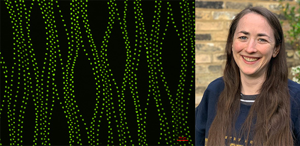
Microscopy Prize
Awarded to Research Associate Dr Niamh Fox, who is from the Fluids in Advanced Manufacturing research group at the Institute for Manufacturing (IfM), part of the Department of Engineering, for her image titled Meshing drug delivery with flexible supports.
Niamh said: “My entry is a magnified fluorescence image of droplets of fluorescein in polyethylene glycol during development of new methods of drug release. During curing, droplets are printed into a polymer used in a wide range of applications called polydimethylsiloxane (PDMS). The balance of surface tension pins the drop at the surface of a curing PDMS film. The top of the droplets are left peeking above the PDMS film. This creates an open pore when it cures, which allows drug release. Convection currents during the curing of PDMS cause the self-organisation of the droplets – originally printed at regular spacings – giving rise to the mesh-like patterns observed, if there isn’t the proper balance between droplet printing and curing time.”
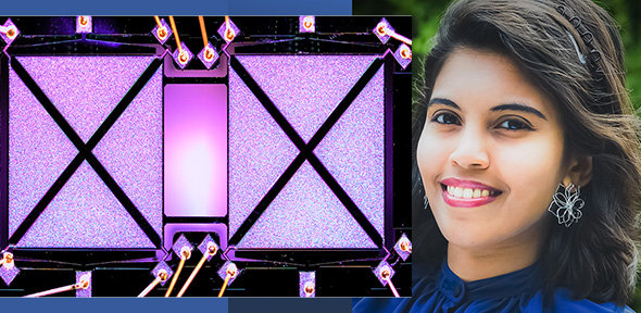
Head of Department Prize
Awarded to Malar Chellasivalingam for her image titled Highly sensitive weakly-coupled MEMS Resonators. At the time of entering the competition, Malar was a PhD student in the Department of Engineering, supervised by Professor Ashwin A. Seshia.
Malar said: “My PhD research work focused on implementing miniaturised sensors, based on MEMS (micro-electro-mechanical systems) technology, for sensing ultra-fine particles that are emitted from various sources such as exhaust plumes in aircraft engines, diesel engines or automobile exhausts, industrial emissions, dust, smoke.
“These ultra-fine particles are less than 100 nanometres in diameter, and research has highlighted the significant risks to human health when these particles are inhaled.
“My microscopic image shows a silicon MEMS resonator designed and fabricated as such that it will be highly sensitive to nano-sized particles when used as mass sensors. It has already shown three orders of magnitude improvement in sensitivity to the nano-sized particles, compared to more conventional gravimetric MEMS sensors of similar dimensions.
“The image shows two square-shaped MEMS resonators, coupled to each other by means of two mechanical beam couplers that enable weak coupling between the square plate resonators. The MEMS resonators are ball-bonded (wire-bonded) on all of its corner points to establish electrical connections to the device.”
The panel of judges included Allan McRobie, Professor of Structural Engineering; Stephen Furzeland from ZEISS; Professor Richard Prager, Head of Department; and Philip Guildford, Chief Operating Officer.
Philip Guildford said: “The images are not only beautiful, but tell remarkable stories about early career engineers at the frontiers of technology, changing the world with their brilliant research.”
- View the shortlisted entries.
- All of the 2020-21 entries can be seen on the Department’s Flickr page.
